This is a demo store. No orders will be fulfilled.

PMA(叠氮溴化丙锭), 20 mM in water

有货

库存信息

关闭
货号 (SKU) 包装规格 是否现货 价格 数量
P598082-100μl
100μL 现货 Stock Image

基本描述

产品名称 PMA(叠氮溴化丙锭), 20 mM in water
产品介绍

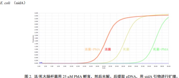
PMA是一种高亲和性的DNA结合染料,该染料本身具有微弱的荧光,但与核酸结合后可以发出更为明亮的荧光。它尤其与双链DNA具有高亲和性。PMA不具有细胞膜渗透性,因此可选择性地修饰膜受损的死细胞的 DNA。PMA修饰的DNA经蓝光(~464 nm)光解后,PMA上的光反应性叠氮基转化为高反应性氮烯自由基,与DNA结合位点附近的任何烃部分反应形成稳定的共价氮碳键,从而导致永久性 DNA 修饰(图1)。该修饰过程会使DNA不溶,并使其在随后的基因组DNA提取过程中与细胞碎片一起丢失,进而阻碍死细胞中目标DNA的PCR扩增。残留在溶液中未结合的PMA,在强光照射下与水分子反应分解成无交联活性的羟胺化合物,使羟胺不再能够共价结合DNA,从而不影响PCR扩增。这个特性使得 PMA 可以通过实时定量 PCR(qPCR)的手段,检测多种样本类型包括:细菌、生物膜、酵母、真菌、病毒和真核细胞;与qPCR、NGS、Sanger测序和LAMP技术相结合,广泛应用在食品和水安全和环境测试中。


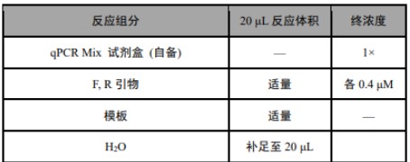
产品参数:

橙红色液体; Ex (pH 3) = 464 nm (before photolysis); Ex /Em (after photocrosslinking to nucleic acid) = 510/610 nm

注意事项:

1. 使用前请将产品瞬时离心至管底,再进行后续实验。 2. 试剂盒组分中含有荧光染料,使用及保存过程中注意避光。 3. 为了您的安全和健康,请穿实验服并戴一次性手套操作。

应用范围:

细菌核酸染色

使用方法:
使用前注意事项:
1. PMA 根据细胞膜通透性区分死菌和活菌。许多杀死细菌的方法会导致细胞膜受损,因此适用于 PMA 检测。 但有些方法,如紫外线照射,可能不会立即导致细胞膜破裂。因此,在选择 PMA 检测之前,需先进行文献检索和预实验确定是否适用于您选择的细菌类型和杀伤方法。
2. PMA 处理完细菌后需要进行光解,以使染料与死细胞 DNA 共价结合。光解操作可以使用蓝色或白色光源。一般来说,灯越亮,光解步骤的效率就越高。非 LED 灯(如卤素灯)可能会加热您的样本并对分析产生负面影响,照射时需要使用冰块对样品降温处理。
3. 样本可以在光解后冷冻保存。若在进行 PMA 处理光解之前冷冻样本可能会损坏细胞膜并产生假阴性结果。如需在检测之前冷冻样本,建议先进行预实验。
4. PMA 的部分作用机制是通过沉淀从样本中去除 PMA 共价修饰的 DNA;因此,在提取基因组 DNA 时,需使用相同体积的基因组 DNA 洗脱液,进行体积归一化处理。阳性对照可以使用活细胞的基因组 DNA。
5. 为了验证 PMA 在测试样本中的有效性,可对比同处理样本-/+PMA 之间的 Ct (dCt)变化。

实验材料(自备):
①光源(用于 PMA 修饰 DNA 后的光解步骤);②细菌基因组 DNA 提取试剂盒;③配套的 qPCR Mix 试剂盒;④对应样本类型的有效 qPCR 引物;
实验步骤:


1. 吸取 10 μL 菌液于液体 LB 培养基,在细菌培养箱中培养过夜或更长时间,使细菌培养至对数生长期(OD600 ≈ 1.0);
注:培养基类型及培养时间根据实验调整。
2. 取活细菌两份,每份 400 μL,置于干净离心管中。
3. (建议)制备死细菌。若需要死细菌作为对照,可将活细菌放入水浴锅中 95℃加热 5 min,或 58℃加热 3 h,具体操作根据样本类型自行选择,即可获得死细菌。死细菌的后续操作同活细菌。
4. 两份活细菌,一份不用 PMA 处理,一份用 25 μM PMA 处理(处理不同类型或不同来源细菌的最适 PMA 浓度需查阅相关文献)。
5. 将 PMA 处理的样本室温置于摇床上,避光孵育 10 min,使染料与样本充分混匀。
6. 将样品曝光,可以使用蓝色或白色光源,照射时间需自行摸索。例如,可用 60 W 的蓝光灯,照射 15 min。
注:① 若使用卤素灯,我们建议将 PMA 处理的样本管放在距离光源 20 cm 处的冰块上。冰块应放在透明托盘中,调整光源使其直接指向样品,光解 5-15 min;② 若直接用环境中获取的细菌进行实验,由于环境样本的复杂性或浑浊度,需适当延长光解时间。

7. 处理和未处理的活细菌 5000 ×g,离心 10 min,去上清。
8. 根据样本类型选择合适的基因组 DNA 提取试剂盒,洗脱 DNA 时,各组样本使用相同的洗脱体积。
注:DNA 提取步骤参考所用试剂盒的说明书。PMA 的部分作用机制是通过沉淀从样品中去除 PMA 结合的 DNA;因此,在提取基因组 DNA 时,各组应使用相同体积的基因组 DNA 洗脱液,进行体积归一化处理(死菌和活菌提取基因组 DNA 量不一致,因此两者浓度相差明显)。
9. 按如下体系制备反应混合液:

注:① 商用 DNA 提取试剂盒提取的 DNA,qPCR 模板以 2 μL 作为起始优化体积;② 模板体积不得超过最终反应体积的10%;③ 模板浓度:gDNA 做模板时,通常 1-10 ng 即可;④ PCR 引物的终浓度通常为 0.4 μM,可以得到较好的结果,反应性能较差时,可以在 0.2 - 1 μM 范围内调整引物浓度。
10. 轻轻涡旋混匀反应混合液,转移固定体积至 PCR 管。
11. 测试程序(根据自备 qPCR Mix 试剂盒程序进行程序设定)。
12. 数据分析
使用活细菌和死细菌作为对照,分析计算样本中活细胞数。建议开始 PMA qPCR 活细菌检测前,先验证引物及 PCR 程序是否合适。死、活细菌对照 dCt 计算
(1) qPCR 结束后,使用仪器软件计算每个样本的 Ct 值。
(2) 通过计算每个对照细菌的 dCt 的方式,判断 PMA 是否成功抑制了死细菌 DNA 的扩增,计算如下:
dCt 活 = Ct(活, PMA 处理) - Ct(活, PMA 未处理)
dCt 死 = Ct(死, PMA 处理) - Ct(死, PMA 未处理)
(3) 活细菌的 dCt 期望值接近于 0 ± 1,这表明 PMA 不会影响活细胞 DNA 的扩增;
(4) 死细菌的 dCt 期望值大于 4(dCt 为 4 表示减少了约 16 倍,即去除了 94% 的死细菌 DNA;dCt 为 8 表示减少了约250 倍,即去除了 99.6%的死细菌 DNA)。
(5) 死细菌的 dCt 取决于许多因素,包括:菌株系/细胞类型;细菌被杀死方式;PMA 使用浓度;扩增序列长度。
13. 活细菌占比计算
如果死、活细菌对照结果正常,可以进入样本中活细菌占比计算。
(1) 计算样本的 dCt 值:
dCt 样本 = Ct(样本, PMA 处理) - Ct(样本, PMA 未处理)
(2) 转换 dCt 值为活细菌比例:
PMA 抑制倍数 = 2(样本 dCt)
活细菌% = 100 / PMA 抑制倍数
14. 计算活细菌的绝对数量
如果想计算样本中活细菌的绝对数量☆,需要用已知数量的目标菌的基因组 DNA 制作标准曲线。建议稀释的几组基因组浓度在 qPCR 分析体系的范围内。
(1) 以 Ct 值为纵坐标,细胞数为横坐标,制作标准曲线。
(2) 计算实验样本的拷贝数:
Ct = 斜率 * 细胞数 + y 轴截距 (y = mx + b)
细菌数样本 = (Ct – y 轴截距) / 斜率
注:纯化过程中活细菌 DNA 未损失。

示例:


PMA is a DNA binding dye with high affinity. The dye itself has weak fluorescence, but it can emit brighter fluorescence after binding with nucleic acids. It especially has high affinity with double stranded DNA. PMA does not have cell membrane permeability, so it can selectively modify the DNA of dead cells with damaged membranes. After bllight (~464 nm) photolysis of PMA modified DNA, the photoreactive azido group on PMA is converted into highly reactive azene radical, which reacts with any hydrocarbon moiety near the DNA binding site to form a stable covalent nitrogen carbon bond, resulting in permanent DNA modification (Figure 1). This modification process will make DNA insoluble and make it lost together with cell debris in the subsequent genomic DNA extraction process, thus hindering the PCR amplification of target DNA in dead cells. The unbound PMA remaining in the solution reacts with water molecules under strong light irradiation and decomposes into hydroxylamine compounds without cross-linking activity, so that hydroxylamine can no longer covalently bind DNA, thus not affecting PCR amplification. This feature enables PMA to detect a variety of sample types including bacteria, biofilms, yeast, fungi, viruses and eukarYOtic cells by real-time quantitative PCR (qPCR); Combined with qPCR, ngs, Sanger sequencing and lamp technology, it is widely used in food and water safety and environmental testing.


Product parameters:

Product parameters:Orange-red liquid; Ex (pH 3) = 464 nm (before photolysis); Ex /Em (after photocrosslinking to nucleic acid) = 510/610 nm

Matters needing attention:

1. please centrifuge the product to the bottom of the tube immediately before use, and then conduct subsequent experiments. 2. the components of the kit contain fluorescent dyes. Avoid light during use and storage. 3. for your safety and health, please wear experimental clothes and disposable gloves.

Scope of application:

Bacterial nucleic acid staining

Instruction:
Precautions before use:
1.PMA distinguishes dead and live bacteria according to cell membrane permeability. Many methods of killing bacteria can cause cell membrane damage, so they are suitable for PMA detection. But some methods, such as ultraviolet irradiation, may not immediately cause cell membrane rupture. Therefore, before selecting PMA detection, it is necessary to conduct literature search and pre-experiment to determine whether it is suitable for the bacterial type and killing method you choose.
2.After PMA treatment, the bacteria need to be photolyzed to covalently bind the dye to dead cell DNA. Photolysis operations can use blue or white light sources. Generally speaking, the brighter the lamp, the higher the efficiency of the photolysis step. Non-LED lamps ( such as halogen lamps ) may heat your sample and have a negative impact on the analysis. Ice is required to cool the sample during irradiation.
3.Sample can be cryopreservation after photolysis. Frozen samples before PMA treatment photolysis may damage the cell membrane and produce false negative results. If the sample needs to be frozen before detection, it is recommended to perform a pre-experiment first.
4.Part of the mechanism of PMA is to remove PMA covalently modified DNA from the sample by precipitation ; therefore, when extracting genomic DNA, it is necessary to use the same volume of genomic DNA eluent for volume normalization. The positive control can use the genomic DNA of living cells.
5.In order to verify the effectiveness of PMA in the test sample, the Ct ( dCt ) changes between- / + PMA can be compared.

Experimental materials ( self-provided ):
①Light source ( used for the photolysis step after PMA modified DNA ) ; ② Bacterial genomic DNA extraction kit ; ③ qPCR Mix kit ; ④ effective qPCR primers corresponding to the sample type ;
Experimental procedure:

1.Suck 10 μL of bacterial liquid in liquid LB medium, culture overnight or longer in the bacterial incubator, so that the bacterial culture to the logarithmic growth phase(OD600 ≈ 1.0)
note : The type of culture medium and culture time were adjusted according to the experiment.
2.Two copies of viable bacteria, 400 μL each, were placed in a clean centrifuge tube.
3. ( Recommended ) Preparation of dead bacteria. If dead bacteria are needed as a control, live bacteria can be placed in a water bath at 95 ° C for 5 min, or heated at 58 ° C for 3 h. The specific operation is self-selected according to the sample type to obtain dead bacteria. The subsequent operation of dead bacteria is the same as that of live bacteria.
4.Two live bacteria, one without PMA treatment, and one with 25 μM PMA treatment ( the optimal PMA concentration for different types or different sources of bacteria needs to be referred to the relevant literature ). The PMA-treated samples were placed on a shaker at room temperature and incubated in the dark for 10 min to fully mix the dye with the sample.
6.Exposure of the sample, you can use blue or white light source, irradiation time to explore their own. For example, a 60 W blue light can be used for 15 min.
Note : 1 If a halogen lamp is used, we recommend that the PMA-treated sample tube be placed on an ice block 20 cm away from the light source. The ice should be placed in a transparent tray, and the light source should be adjusted to point directly to the sample for photolysis for 5-15 min. If the bacteria obtained from the environment are directly used for experiments, due to the complexity or turbidity of the environmental samples, the photolysis time needs to be prolonged appropriately.
7.Treated and untreated live bacteria 5000 × g, centrifuged for 10 min, and the supernatant was removed.
8.Select the appropriate genomic DNA extraction kit according to the sample type, and use the same elution volume for each group of samples when elution DNA.
Note : DNA extraction steps refer to the instructions of the kit used. Part of the mechanism of action of PMA is to remove PMA-bound DNA from the sample by precipitation ; therefore, when extracting genomic DNA, each group should use the same volume of genomic DNA eluent for volume normalization ( the amount of genomic DNA extracted from dead bacteria and live bacteria is inconsistent, so the concentration of the two is significantly different ).
9.Preparation of reaction mixture according to the following system :

 
Note : 1 For the DNA extracted by commercial DNA extraction kit, the qPCR template was optimized with 2 μL as the initial volume ; 2 The template volume should not exceed 10 % of the final reaction volume ; 3 Template concentration : gDNA as template, usually 1-10 ng ; the final concentration of PCR primers is usually 0.4μM, which can get better results. When the reaction performance is poor, the primer concentration can be adjusted in the range of 0.2-1μM.
10.Slightly vortex the reaction mixture, transfer the fixed volume to the PCR tube.
11. Test procedures ( according to the own qPCR Mix kit program to set the program ).
12. Data analysis
Using live bacteria and dead bacteria as controls, the number of live cells in the sample was analyzed and calculated. It is recommended to verify the suitability of primers and PCR procedures before starting PMA qPCR detection of live bacteria.
Calculation of dead and living bacteria control dCt
( 1 ) After qPCR, the Ct value of each sample was calculated using the instrument software.
( 2 ) By calculating the dCt of each control bacteria, it was judged whether PMA successfully inhibited the amplification of dead bacterial DNA. The calculation is as follows :
dCt live = Ct ( live, PMA treated ) -Ct ( live, PMA untreated )
dCt die = Ct ( die, PMA treated ) -Ct ( die, PMA untreated )
( 3 ) The expected value of dCt of living bacteria was close to 0 ± 1, which indicated that PMA did not affect the DNA amplification of living cells.
( 4 ) The expected value of dCt of dead bacteria is greater than 4 ( dCt is 4 means that it is reduced by about 16 times, that is, 94 % of dead bacterial DNA is removed ; the dCt of 8 represents a reduction of about 250 times, that is, 99.6 % of the dead bacterial DNA is removed ).
( 5 ) The dCt of dead bacteria depends on many factors, including : strain / cell type ; the way bacteria are killed ; the concentration of PMA used ; amplified sequence length.
13.Calculation of the proportion of live bacteria
If the control results of dead and live bacteria are normal, the proportion of live bacteria in the sample can be calculated.
( 1 ) Calculate the dCt value of the sample :
dCt sample = Ct ( sample, PMA treated ) -Ct ( sample, PMA untreated )
( 2 ) Conversion of dCt value to live bacteria ratio :
PMA inhibition multiple = 2 ( sample dCt )
Viable bacteria % = 100 / PMA inhibition multiple
14. Calculating the absolute number of viable bacteria
If you want to calculate the absolute number of viable bacteria in the sample, you need to use a known number of target bacteria genomic DNA to make a standard curve. It is recommended that the diluted concentrations of several groups of genomes are within the range of the qPCR analysis system.
( 1 ) The standard curve was made with the Ct value as the ordinate and the cell number as the abscissa.
( 2 ) Calculate the copy number of the experimental samples :
Ct = slope * cell number + y axis intercept ( y = mx + b )
Bacterial count sample = ( Ct-y axis intercept ) / slope
Note : The live bacterial DNA was not lost during the purification process.
Examples :


储存与运输

储存温度 避光,-20°C储存
运输条件 超低温冰袋运输
CAS编号和信息 P598082
分子类型 未知

质检证书(CoA,COO,BSE/TSE 和分析图谱)

C of A & Other Certificates(BSE/TSE, COO):
输入批号以搜索分析图谱:

通过匹配包装上的批号来查找并下载产品的 COA,每批产品都进行了严格的验证,您可放心使用!

找到8个结果

批号(Lot Number) 证书类型 货号
H2519405 分析证书 P598082
H2519404 分析证书 P598082
H2519403 分析证书 P598082
C2528458 分析证书 P598082
J2429537 分析证书 P598082
F2417211 分析证书 P598082
F2417212 分析证书 P598082
J2318706 分析证书 P598082

技术文档和文章

蛋白质印迹通用实验方案
General Protocol for Western Blotting
蛋白质印迹的裂解物制备法
Methods of Lysate Preparation for Western Blotting
General Protocol for Western Blotting
Methods of Lysate Preparation for Western Blotting
荧光标记抗体的蛋白质印迹法
Western Blotting with Fluorescently Labeled Antibodies
蓝色天然聚丙烯酰胺凝胶电泳实验方案 (BN-PAGE)
BN-PAGE Protocol
BN-PAGE Protocol
Western Blotting with Fluorescently Labeled Antibodies
General Protocol for Western Blotting
组蛋白的蛋白质印迹法
Histone Western Blot
蛋白质去磷酸化实验方案
Protein Dephosphorylation Protocol

溶液计算器