This is a demo store. No orders will be fulfilled.
 首页
首页 400-620-6333
Annexin V staining
Product Description
Annexin V is a calcium-dependent phospholipid-binding protein with high affinity for phosphatidylserine (PS) exposed on the outer leaflet of the plasma membrane. In healthy cells, PS is restricted to the inner leaflet, but during apoptosis, PS translocates to the cell surface, serving as a key marker of apoptosis. Fluorescently or otherwise labeled Annexin V enables sensitive detection of apoptotic cells in the presence of Ca²⁺, and is widely used in flow cytometry, fluorescence microscopy, and various apoptosis assays.
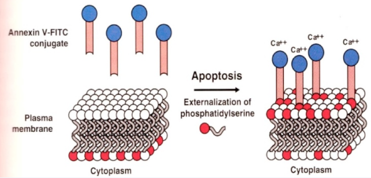
Figure 1. Schematic of Phosphatidylserine (PS) Externalization During Apoptosis
Why Use Annexin V to Detect Apoptosis?
Annexin V enables early detection of apoptotic cells by recognizing the externalization of phosphatidylserine (PS) before morphological changes occur. With nanomolar affinity for PS, it can accurately distinguish apoptotic from viable cells. Annexin V is widely applicable to flow cytometry, microscopy, and other analysis platforms, supporting both quantitative measurement and visualization of PS distribution.
Annexin V Conjugates for Apoptosis Detection
- Sensitive detection, precise differentiation of apoptosis stages: By combining with PI, 7-AAD, or DAPI, early and late apoptotic cells can be accurately identified.
- Multiple conjugate options, broad compatibility: A variety of labels, including AF405, AF488, FITC, PE, APC, etc., are available, compatible with different laser systems.
- Simple operation, flexible application: Can be used alone or in combination with nuclear dyes, suitable for flow cytometry and fluorescence microscopy analysis.
Product List
|
Annexin V Staining Experimental Conditions
Annexin V staining is preferably performed on unfixed, live cells or tissues. If the samples have been fixed, specific conditions are required to preserve signal intensity: Annexin V staining should be performed first in the presence of Ca²⁺, followed by short-term fixation (10–15 minutes) with 1% paraformaldehyde (PFA) in Ca²⁺-containing buffer. Alcohol- or methanol-based fixation, as well as any treatments involving detergents or permeabilization, should be avoided.
To enhance staining performance, we provide Annexin V Binding Buffer (A1372288), which optimizes the interaction between Annexin V and phosphatidylserine (PS). This buffer offers the following advantages:
- Promotes specific binding: The buffer contains an optimized concentration of calcium ions, which effectively facilitates the binding of Annexin V to phosphatidylserine (PS) on the cell surface.
- Suitable for early apoptosis detection: Provides essential conditions for detecting early-stage apoptosis by flow cytometry, enhancing sensitivity and accuracy.
Applications of Annexin V Staining
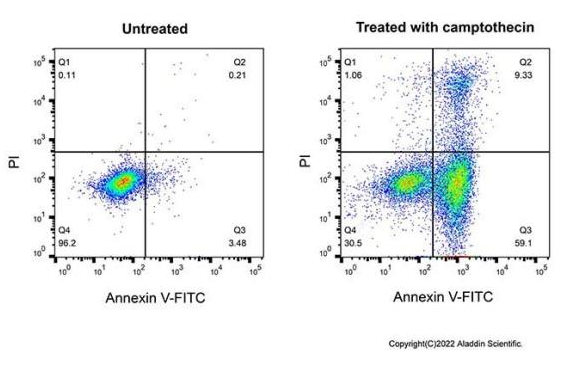
Flow Cytometry: Annexin V-FITC (rp226053)
U937 cells were either left untreated (left) or treated (right) with 4µM camptothecin (C111281) for 4 hours, then stained with Annexin V-FITC (rp226053) and propidium iodide (PI) (P1372285) in Annexin V Binding buffer (A1372288) for 10 minutes at room temperature.

Flow Cytometry: Annexin V-PE (rp226055)
U937 cells were either untreated (blue) or treated (red) with 5µM camptothecin (C111281) for 4 hours, then stained with Annexin V-PE (rp226055) in Annexin V Binding buffer (A1372288) for 10 minutes at room temperature.
Cell Apoptosis Detection Kit
In early apoptosis, the cell membrane remains intact, preventing viability dyes such as Propidium Iodide (PI) or 7-AAD from entering the cell. As a result, these cells are stained only by Annexin V and not by the viability dye, allowing for the identification of early apoptotic cells. As apoptosis progresses to the late stage, loss of membrane integrity enables Annexin V to bind intracellular phosphatidylserine (PS), and viability dyes can also enter the cell. By combining Annexin V with a viability dye, early apoptotic cells (Annexin V positive, viability dye negative) can be distinguished from late apoptotic or necrotic cells (Annexin V positive, viability dye positive).
Our kits are specifically designed for flow cytometry and allow accurate identification and quantification of dead cells, including both apoptotic and necrotic populations.
Product List
Name | Kit Components | Packaging | Catalog Number |
Annexin V-FITC/PI Apoptosis Detection Kit | 10× Annexin V Binding Buffer;Annexin V-FITC;Propidium iodide Staining Solution (PI) | 20T/50T/ 100T | |
Annexin V-APC/PI Apoptosis Detection Kit | 10× Annexin V Binding Buffer;Annexin V (APC);Propidium iodide Staining Solution (PI) | 20T/50T/ 100T | |
Annexin v-pe / rednucleus Ⅱ apoptosis Kit
| 1×Annexin V Binding Buffer;Annexin V-PE;RedNucleus II | 10T/50T/ 100T | |
Annexin V-FITC/PI Apoptosis Detection Kit | 4×Annexin V Binding Buffer;Annexin V-FITC;Propidium Iodide, PI | 20T/50T/ 100T |
Applications of Cell Apoptosis Detection Kits
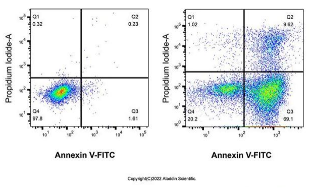
Annexin V-FITC/PI Apoptosis Detection Kit (A1372286) - Flow Cytometry
Flow cytometric analysis of U937 cells untreated (left) or treated with 4µM camptothecin (C111281) for 4 hours using Annexin V-FITC/PI Apoptosis Detection Kit (A1372286).

Annexin V-APC/PI Apoptosis Detection Kit (A1372287) - Flow Cytometry
Flow cytometric analysis of U937 cells untreated (left) or treated with 4µM camptothecin (C111281) for 4 hours using Annexin V-APC/PI Apoptosis Detection Kit (A1372287).
Aladdin: https://www.aladdinsci.com/
Categories: 技术文章