
Determine the necessary mass, volume, or concentration for preparing a solution.
This is a demo store. No orders will be fulfilled.
| SKU | Size | Availability |
Price | Qty |
|---|---|---|---|---|
|
rp155876-0.1ml
|
0.1ml |
≥10
|
$19.90
|
|
|
rp155876-0.5ml
|
0.5ml |
5
|
$59.90
|
|
|
rp155876-1ml
|
1ml |
5
|
$99.90
|
|
Streptavidin was conjugated with FITC under optimum conditions, and unconjugated Streptavidin and free FITC were removed.
| Product Name | Streptavidin protein (FITC)used for Flow Cytometry; IF; various applications |
|---|---|
| Synonyms | FITC Streptavidin | Streptavidin-FITC | FITC-streptavidin conjugate | SAv-FITC | Streptavidin Fluorescein Conjugated | Streptavidin-Fluorescein Isothiocyanate | SA-FITC | SA (FITC) | SA V1 | SA V2 | strepavidin | Streptavidin V1 | Streptavidin V2 |
| Grade | Azide Free, Em:517nm, Ex:498nm |
| Product Description |
Background Streptavidin is a tetrameric bacterial protein isolated from Streptomyces avidinii providing 4 high-affinity biotin binding sites. Streptavidin homo-tetramers have an extraordinarily high affinity for biotin. With a dissociation constant on the order of ≈10⁻¹⁴ mol/L, the binding of biotin to streptavidin is one of the strongest non-covalent interactions known in nature. Unlike egg-white avidin, which has a net positive charge at neutral pH and contains about 7% carbohydrate, streptavidin has almost no net charge at neutral pH, does not contain carbohydrate, and exhibits lower non-specific background. Streptavidin conjugates are widely used together with a conjugate of biotin for specific detection of a variety of proteins, protein motifs, nucleic acids and other molecules. This FITC-streptavidin conjugate was prepared by highly purified Streptavidin and free FITC was removed. Streptavidin (FITC) is a useful second-step reagent for the indirect immunofluorescent staining of cells in combination with biotinylated primary antibodies for flow cytometric analysis. Excitation at 488nm light leads to a fluorescence emission maximum of 520 nm. Recommended Usage: Every lot of Streptavidin-FITC is tested by flow cytometry using biotinylated primary antibodies. From this testing it is recommended that between 0.02 and 0.25 µg of streptavidin be used per 106 cells in a 100 µl staining volume. |
| Specifications & Purity | Azide Free, Ex:498nm, Em:517nm, Streptavidin Concentration: 1 mg/mL |
| Protein Length | Full length protein |
| Conjugation | FITC |
| Excitation(nm) | Blue Laser(488nm) |
| Emission(nm) | 517nm |
| Ex/Em(nm) | |
| Source | Native |
| Application | Flow Cytometry; IF; |
|---|---|
| Concentration | Streptavidin Concentration: 1 mg/mL |
| Storage Temp | Store at 2-8°C,Avoid repeated freezing and thawing |
| Shipped In | Wet ice |
| Stability And Storage | Shipped at 4℃. Store at 4°C and protected from prolonged exposure to light. Do not freeze. |
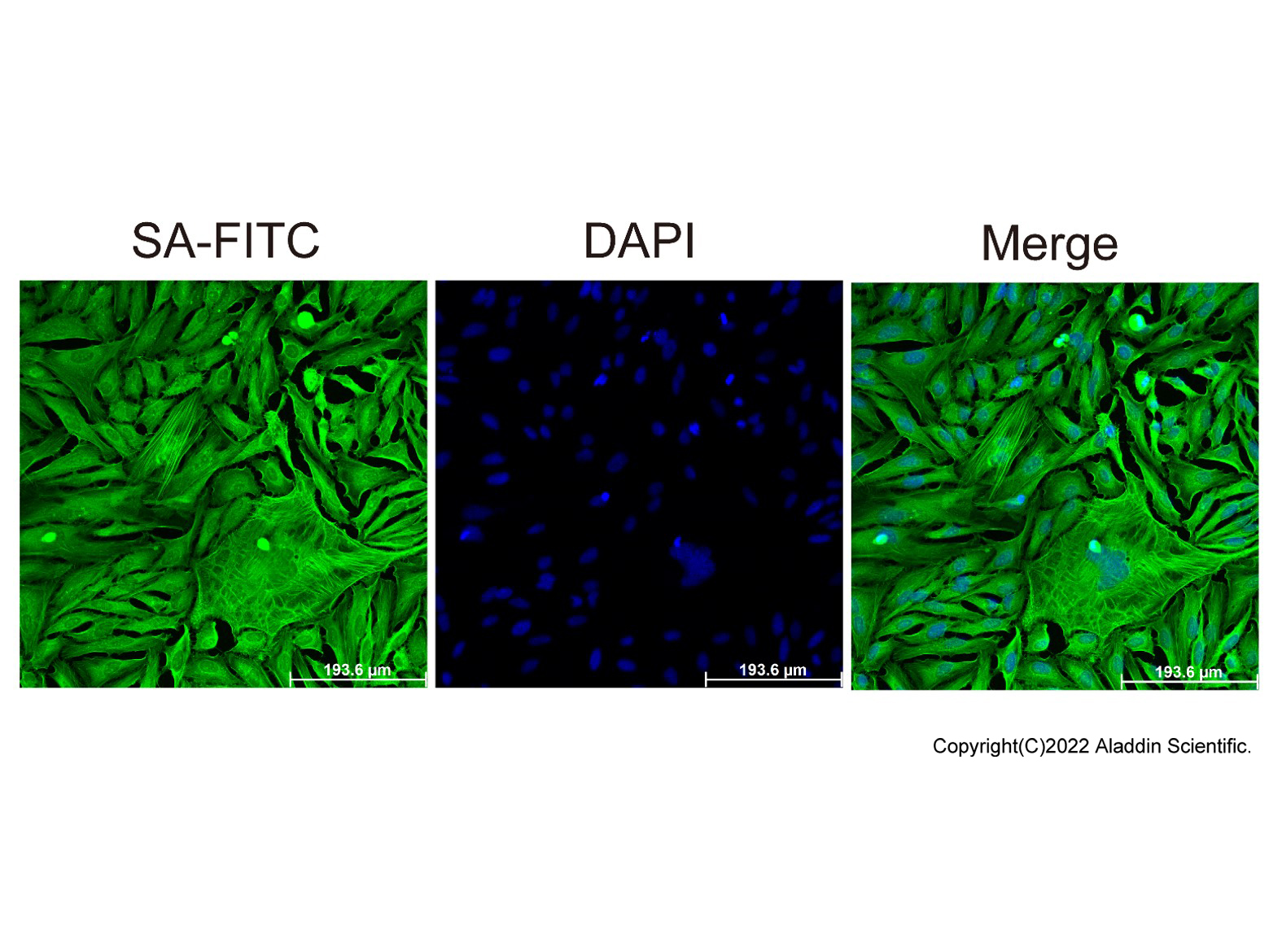
ICC/IF-Streptavidin protein (FITC) (rp155876)
ICC/IF with Streptavidin protein (FITC) (rp155876) stained HeLa cells. The cells were methanol fixed (5 min) and blocked with 1% BSA . The cells were then incubated with Biotin-γ-actin antibody overnight at 4°C. Then Streptavidin protein (FITC) rp155876 (green) was used at 1:1000 for 1h. DAPI was used to stain the cell nuclei (blue) .

Flow-Streptavidin protein (FITC) (rp155876)
BALB/c splenocytes stained with Biotin-CD45R antibody, followed by Streptavidin protein (FITC) (rp155876) at 1/2500 dilution.
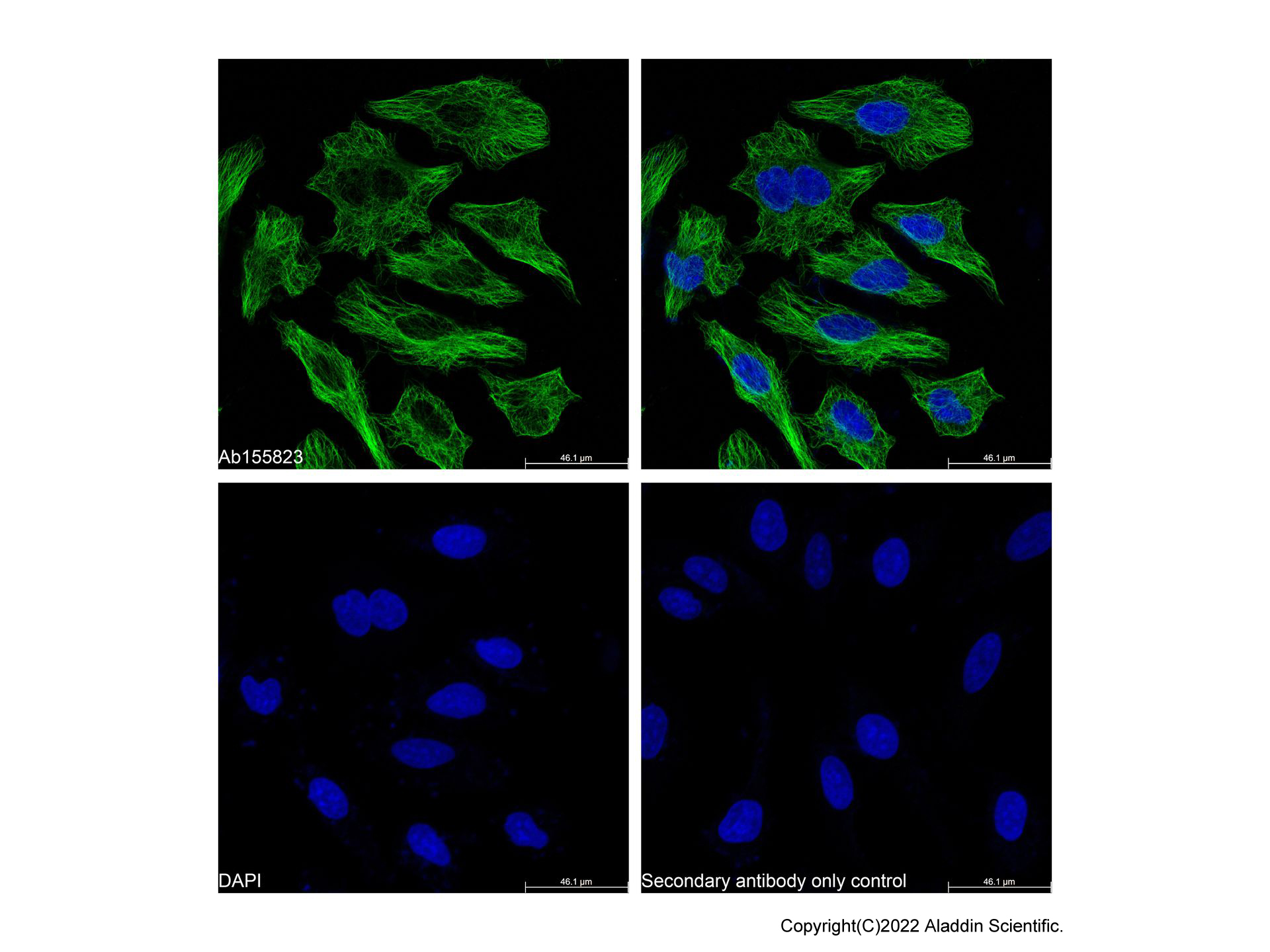
Streptavidin Protein (FITC) (rp155876) - IF
IF analysis of beta Tubulin (green) in HeLa cells. The cells were fixed and permeabilized with 100% methanol for 5 minutes, and blocked with 2% BSA for 1 hour at room temperature. Cells were stained with beta Tubulin Mouse mAb (Ab155823) at 1/2000 dilution in blocking buffer overnight at 4°C, and then incubated with Goat Anti-Mouse IgG H&L (Biotin) (Ab179002) at a dilution of 1/1000 for 1 hour at room temperature in the dark. Then incubated with Streptavidin Protein (FITC) (rp155876) at a dilution of 1/1000 for 1 hour at room temperature in the dark (green). Cells were counterstained with DAPI (blue). PBS instead of the primary antibody was used as the secondary antibody only control. Images were taken on the confocal laser scanning microscope.
| Application | Dilution info |
|---|---|
| Flow Cytometry | 1:500-1:5000 |
| IF/ICC | 1:50-1:500 |
Find and download the COA for your product by matching the lot number on the packaging.
| Lot Number | Certificate Type | Date | Item |
|---|---|---|---|
| Certificate of Analysis | Dec 20, 2023 | rp155876 | |
| Certificate of Analysis | Dec 20, 2023 | rp155876 | |
| Certificate of Analysis | Dec 20, 2023 | rp155876 | |
| Certificate of Analysis | Mar 28, 2023 | rp155876 | |
| Certificate of Analysis | Mar 28, 2023 | rp155876 | |
| Certificate of Analysis | Mar 28, 2023 | rp155876 |